Glaucoma Diagnosis
The only sure way to diagnose glaucoma is with a complete eye exam. A glaucoma screening that only checks eye pressure is not enough to find glaucoma.
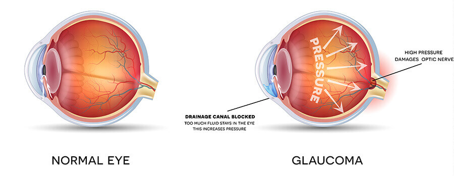
One of the problems with glaucoma, especially open-angle glaucoma, is that there are typically no symptoms in the early stages. Many people who have the disease do not know they have it. This is why it is important to have regular medical eye exams by an ophthalmologist especially as you get older.
Glaucoma Tests
Your ophthalmologist will do the following tests and exams during a comprehensive glaucoma evaluation:
Measure the pressure in your eye (tonometry)
Your doctor measures your eye pressure using tonometry. (See photo above) Testing your eye pressure is an important part of a glaucoma evaluation. A high pressure reading is often the first sign that you have glaucoma. During this test, your eye is numbed with eye drops. Your doctor uses an instrument called a tonometer to measure eye pressure. The instrument measures how your cornea resists pressure. Normal eye pressure generally ranges between 10 and 21 mmHg. However, people with normal-tension glaucoma can have damage to their optic nerve and visual field loss even though their eye pressure remains consistently lower than 21 mmHg.
Inspect your eye’s drainage angle (gonioscopy)
Gonioscopy allows your ophthalmologist to get a clear look at the drainage angle to determine the type of glaucoma you may have. Your ophthalmologist is not able to see your eye’s drainage angle by looking at the front of your eye. However, by using a mirrored lens, he or she can examine the drainage angle to determine if you have open-angle glaucoma (where the drainage angle is not working efficiently enough), closed-angle glaucoma (where the drainage angle is at least partially blocked), or a dangerously narrow-angle (where the iris is so close to the eye’s drain that the iris could block it).
Inspect your optic nerve (ophthalmoscopy)
Your ophthalmologist inspects your optic nerve for signs of damage using an ophthalmoscope, an instrument that magnifies the interior of the eye. Your pupils will be dilated (enlarged) with eye drops to allow your doctor a better view of your optic nerve.
A normal optic nerve is made up of more than one million tiny nerve fibers. As glaucoma damages the optic nerve, it causes the death of some of these nerve fibers. As a result, the appearance of the optic nerve changes. This is referred to as cupping. As the cupping increases, blank spots begin to develop in your field of vision.
Test your side, or peripheral, vision (visual field test)
The visual field test will check for blank spots in your vision. The results of the test show your ophthalmologist if and where blank spots appear in your field of vision — including spots you may not even notice.
The test is performed using a bowl-shaped instrument called a perimeter. When taking the test, a patch is temporarily placed on one of your eyes so only one eye is tested at a time. You will be seated and asked to look straight ahead at a target. The computer makes a noise and random points of light will flash around the bowl-shaped perimeter, and you will be asked to press a button whenever you see a light with your side vision. You should not turn your eyes to look for the lights. Not every noise is followed by a flash of light. Visual field testing is usually performed every 6 to 12 months to monitor for change.
Measure the thickness of your cornea — the clear window at the front of the eye (pachymetry)
Because the thickness of the cornea can affect eye pressure readings, pachymetry is used to measure corneal thickness. A probe called a pachymeter is gently placed on the cornea to measure its thickness. A very thin cornea may increase your risk of glaucoma.